Equations:
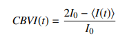
The brain blood volume is extracted from the camera images by calculating the ratio of the mean time-varying intensity ⟨I(t)⟩ over the baseline intensity, cerebral blood volume index CBVI
The brain blood flow is extracted from the camera images by calculating the cerebral blood flow index (CBFI)
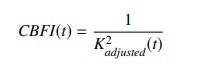
K 2 adjusted is the adjusted speckle contrast (which indicates information about the particle motion, i.e. blood flow velocity in a region) obtained by subtracting the raw speckle contrast K 2 raw by all sources of noise.
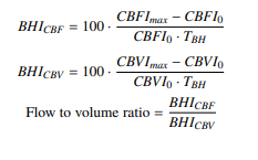
a breath-holding index
Method:
When coherent laser light from a laser diode is directed onto a tissue (e.g., the brain), the light scatters as it passes through the tissue.
The properties of the speckle pattern (a grainy image that appears when a laser illuminates biological tissue) depend on the movement of particles within the tissue, blood cells.The faster the blood flow, the quicker the speckle pattern fluctuates.
where there is no blood flow, the speckle pattern remains relatively constant.
By analyzing the rate at which the speckle pattern changes, SCOS measures the relative speed of blood flow in the tissue. The higher-risk group exhibited a greater increase in brain blood volume.
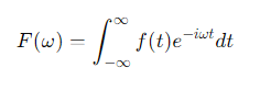
Blood flow signal is recorded as a function of time. Here the heart rate is extracted from the blood flow via Fourier transform.
0 comments:
Post a Comment